


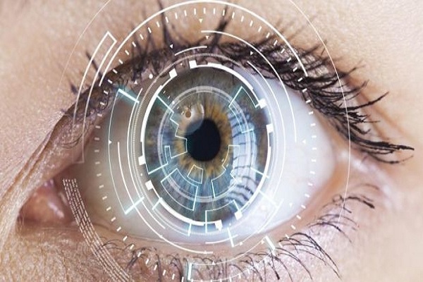
Researchers have used artificial intelligence (AI) to develop a more accurate and detailed method for analyzing images of the back of the eye, an advance that can help ophthalmologists better detect and track eye diseases like glaucoma, and age-related macular degeneration. In the study, published in the journal Scientific Reports, the researchers looked for a new method of analyzing images from a state-of-the-art instrument called the Optical Coherence Tomography (OCT).
The researchers, including those from The Queensland University of Technology (QUT) in Australia, explored a range of machine learning techniques to analyze OCT images.
They tried extracting images from two main tissue layers at the back of the eye from the retina and the choroid. OCT, a commonly used by optometrists and ophthalmologists, takes cross-sectional high-resolution images of the eye, showing different tissue layers.
These images, the study noted, are of tissues about four microns thick. To put that in perspective, the human hair is about 100 microns thick, the researchers said.
Also read: Artificial Intelligence Replacing the Human Effort by 4 times
“The choroid is the area between the retina and the sclera, and it contains the major blood vessels that provide nutrients and oxygen to the eye,” Alonso-Caneiro said. The standard imaging processing techniques used with OCT , he added, defined and analysed the retinal tissue layers well, but very few clinical OCT instruments had the software that analysed choroidal tissue.
According to Alonso-Caneiro, the new method could provide a way to better map and monitor changes in the choroid tissue, and potentially diagnose eye diseases earlier.
Be a part of Elets Collaborative Initiatives. Join Us for Upcoming Events and explore business opportunities. Like us on Facebook , connect with us on LinkedIn and follow us on Twitter.
"Exciting news! Elets technomedia is now on WhatsApp Channels Subscribe today by clicking the link and stay updated with the latest insights!" Click here!